X-ray Microscope Gallery
Sort our gallery by your application or view all images.
If you would like to learn more about our publications and research on specific applications (e.g., batteries), please use the Applications Tab above to navigate to the correct page.
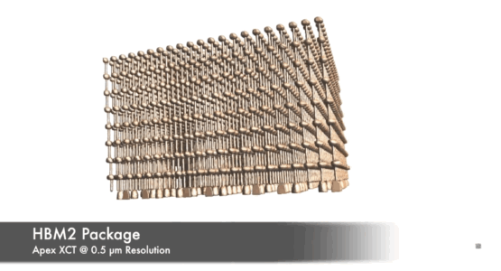
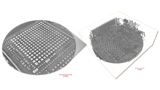
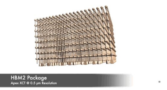
MI25 graphics accelerator board (HBM2 package) at 0.5 µm, imaged with Apex XCT
MI25 graphics accelerator board (HBM2 package) at 0.5 µm, imaged with Apex XCT
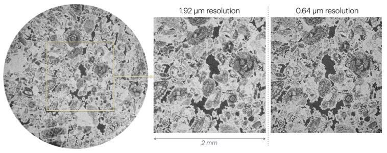
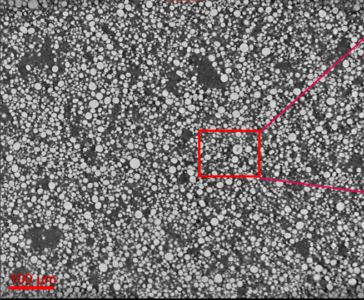
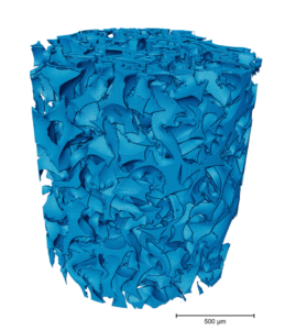
Ocala Limestone
Carbonate rock imaged at different resolutions with EclipseXRM to show detail differences. Courtesy of Prof. Charlotte Garing, UGA.
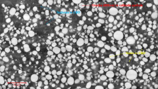
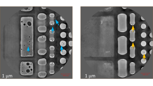
Multiple feature identifications
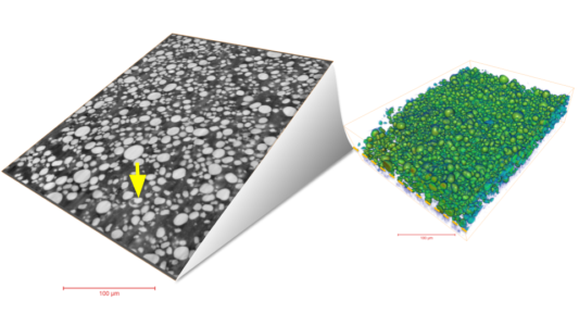
Multiple feature identifications imaged with EclipseXRM on Li-ion battery cathode
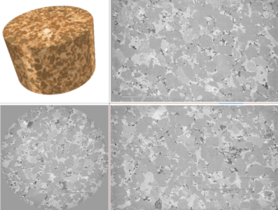
Interior tomography of a sandstone rock
Interior tomography of a 3mm sandstone rock imaged at 0.45um on EclipseXRM
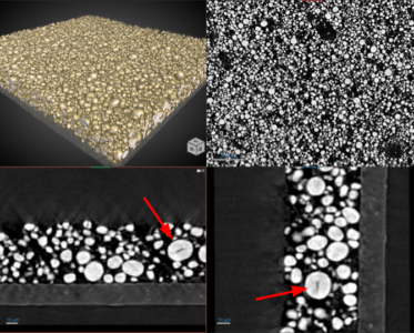
Battery cathode with LiLaZrTAO particles
Battery cathode with LiLaZrTAO particles imaged at 0.25um with EclipseXRM
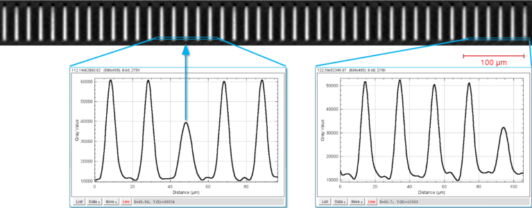
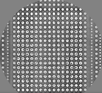
TSVs imaged in 6 minutes
TSVs imaged at 0.3um voxel, acquired within 6 minutes on a 300mm wafer - Imaged on Apex XCT
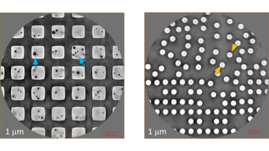
iPhone 14 Pro Region 2 Cracks and Voids
IPhone 14 Pro Region 2 Cracks and Voids Detected at 1µm Voxel - Imaged on Apex XCT
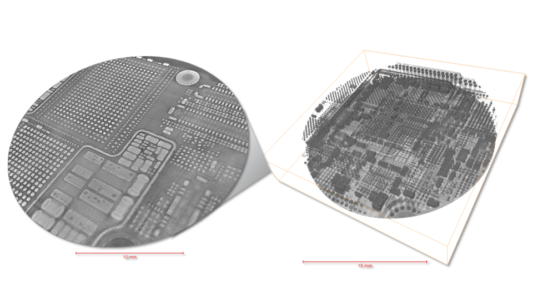
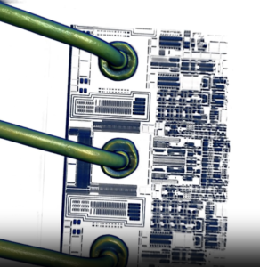
3D rendering of iPhone 14 Pro
3D rendering of iPhone 14 Pro at 8µm voxel at LFOV - Imaged on Apex XCT

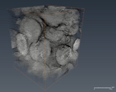
NMC Cathode - 3D Rendering
NMC Cathode - 3D Rendering
Sample courtesy of Dr. Ming Tang, Rice University - Imaged on EclipseXRM

NMC Cathode - 3D Rendering
NMC Cathode - 3D Rendering
Sample courtesy of Dr. Ming Tang, Rice University - Imaged on EclipseXRM
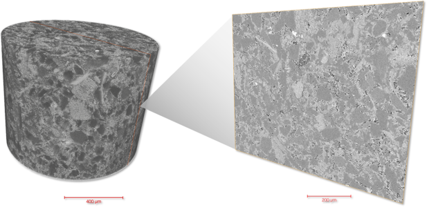
Carbonate Rock
Carbonate rock imaged at 0.322 um, showing detailed mineralogy and pore structures - imaged on EclipseXRM
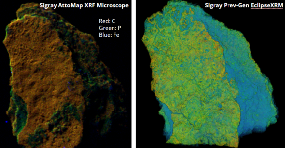
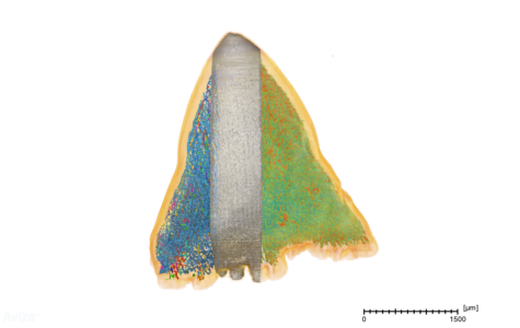
Correlative-calcite microscopy
Correlative microscopy using AttoMap and EclipseXRM to determine Calcium, Phosphorus, and Iron distributions - Imaged on AttoMap and EclipseXRM
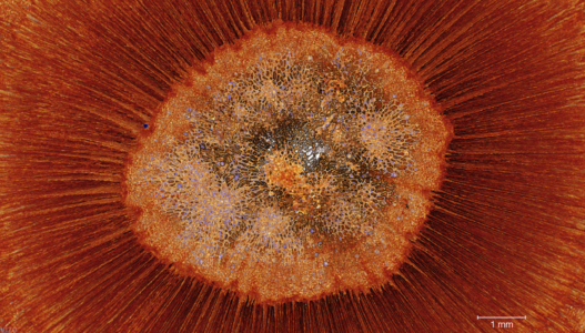
Arabidopsis_Root_3D-XRM_Micrograph
This segment showcases an image of a mutated Arabidopsis stem captured through advanced 3D X-ray computed tomography using the Sigray EclipseXRM X-Ray Microscope.
Sample courtesy of Prof. Breeanna Urbanowicz and Prof. Charlotte Garing, University of Georgia (Athens, GA, USA) - Imaged on EclipseXRM
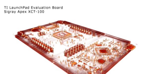
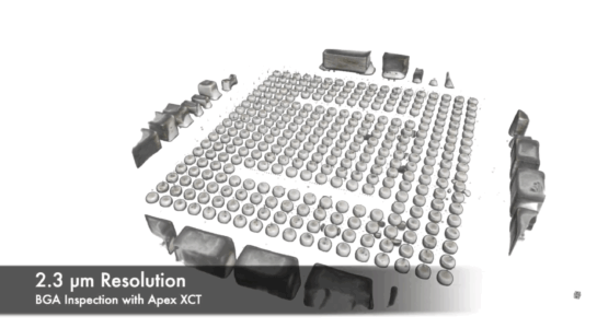
Texas Instruments LaunchPad Evaluation Board
Commercial board scanned at multiple FOVs and voxel sizes: 8 um LFOV (board-level), zooming in at 4 um (component-level), and finally at 1.5 um. LFOV required 20 volumes stitched together and was achieved at only 36 minute per scan. - Imaged on Apex XCT
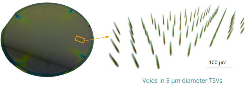
Voids in 5 um TSVs
Voids clearly rendered in 5 um TSVs within minutes. Imaged within an intact 300mm wafer on the Apex XCT.
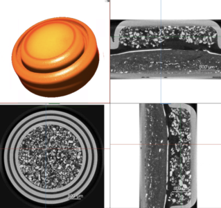
EclipseXRM Coin Cells
Intact coin cells imaged with EclipseXRM, showing system versatility for imaging intact batteries without disassembling
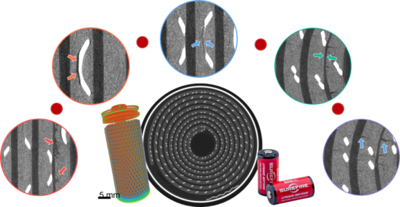
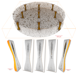
Battery Defects
Zoom-ins show range of defects, include delamination, cracking, pores, misalignments, etc. in an intact commercial battery. A 3D rendering of the battery is seen on the left bottom and the overall scan is shown center. Imaged using the PrismaXRM-810.
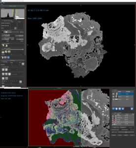
AI-based Segmentation of Kidney Stone
AI-based segmentation of a kidney stone sample acquired on previous generation EclipseXRM to separate different mineral phases.

Pharmaceutical - Compound Houttuynia Cordata 2
Pharmaceutical tablet sample imaged at 7um and then zoomed in at 1um. Houttuynia cordata, also known as Fish Mint or Chameleon Plant, is a medicinal and edible herb containing compounds like volatile oils, flavonoids, and alkaloids, with traditional uses in Asia for conditions like pneumonia, and hypertension. Courtesy of Dr. Guibin Zan.

Pharmaceutical - Compound Houttuynia Cordata
Pharmaceutical tablet sample imaged at 1um. Houttuynia cordata, also known as Fish Mint or Chameleon Plant, is a medicinal and edible herb containing compounds like volatile oils, flavonoids, and alkaloids, with traditional uses in Asia for conditions like pneumonia, and hypertension. Courtesy of Dr. Guibin Zan.
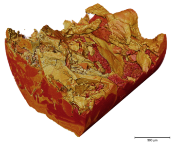
Pathologic human-derived calcite
Pathologic human-derived calcite imaged to show mineralogical differences at high resolution. Imaged using PrismaXRM. Courtesy UCLA Health.
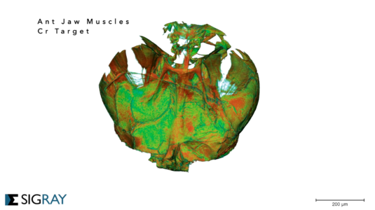
Ant Jaw
Rendering of an unstained ant mandible. Imaged using Sigray's patented multi-spectral source (MSS). Achievable using a ChromaXRM or a PrismaXRM with a MSS option.
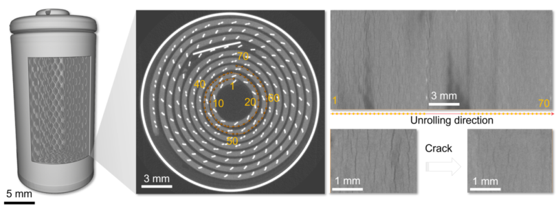
Battery Crack
Cracks in an intact battery electrode. Cracking is more severe in the more tightly rolled portion of the electrode. Imaged using the PrismaXRM-810.
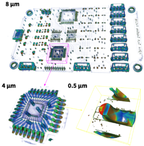
Intact PCB at Multiple Resolutions
Intact PCB imaged to cover the full PCB (8 um voxels) and down to 0.5 um voxels. Imaged on the Apex XCT 150.
J. True et al. Revisiting 3D-X-ray for Rapid Reverse Engineering in Large Electronic Packages and PCBs, PAINE 2022
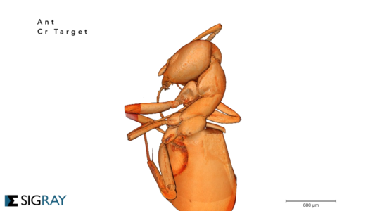
Ant Body
Unstained ant body imaged at 0.7 um resolution using Sigray's patented multi-spectral source (MSS). Achievable using a ChromaXRM or a PrismaXRM with a MSS option.
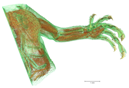
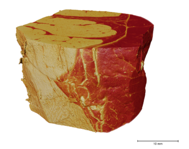
Lizard limb
Lizard limb imaged at 2.1 micron voxel. Outer skin, musculature, and bones are rendered.
Courtesy of Prof. Doug Menke & Prof. Charlotte Garing, University of Georgia Athens - Imaged on PrismaXRM
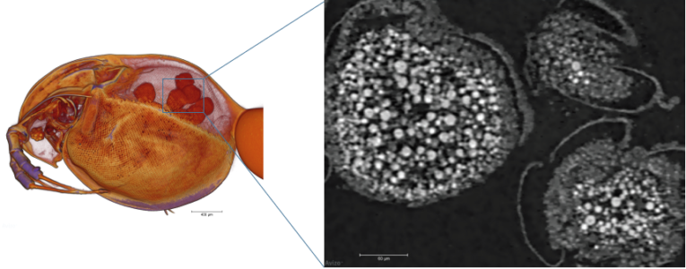
Daphnia Waterflea - ChromaXRM
Unstained daphnia waterflea imaged on ChromaXRM. Eggs can be clearly seen in the sacs of the daphnia.
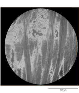
SiC Sample - ChromaXRM
Difficult SiC sample that typically produces very low contrast with microCT. Sigray's ChromaXRM was required.
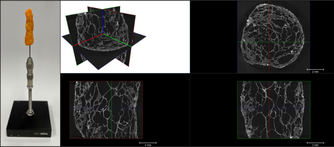
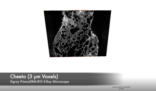
Food Science - Cheetos
Cheetos at high resolution, showing pore space and oil within the pore space. Imaged using PrismaXRM-810.
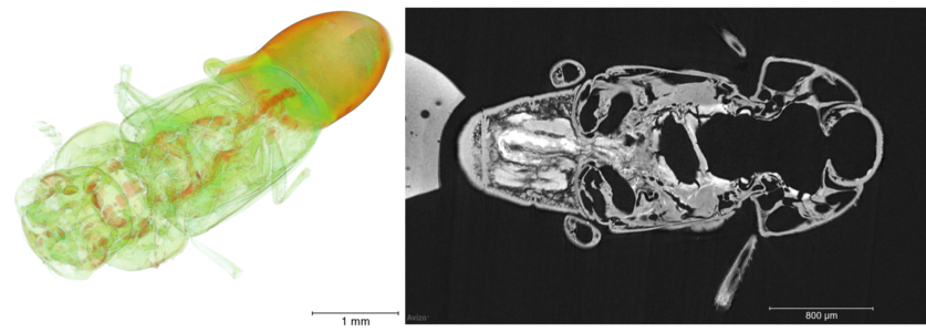
Beetle with Si Nanoparticles
Beetle with Si Nanoparticles imaged on ChromaXRM, showing excellent contrast.
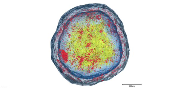
Pores in pharmaceutical pill
Pore network on a polymer coating for a pharmaceutical pill segmented in red. Due to the difficult contrast, this was imaged on ChromaXRM.
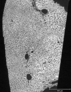
Virtual Histology - ChromaXRM
Virtual histology of an unstained liver sample, showing hepatocyte structure. Imaged on a ChromaXRM.
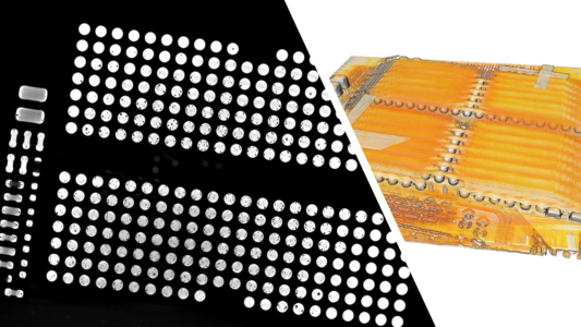
2D virtual slice and 3D rendering of SSD
Bump voids detected on SSD. Shown are a 2D virtual slice taken from a 3D reconstruction. Imaged on a PrismaXRM.

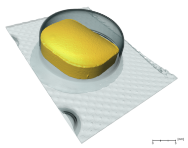
Tylenol pharmaceutical packaging
Package containing Tylenol pills. High resolution of the PrismaXRM reveals cracks in the pills.

